About Our Diagnostic Imaging Service
The diagnostic imaging service provides a comprehensive range of imaging modalities to meet the needs of all our patients, large and small. These include radiography, ultrasound, computed tomography (CT) and magnetic resonance imaging (MRI).
Computed Tomography (CT)
CT imaging is an advanced form of radiography – a more sensitive imaging modality compared to conventional radiographs, that is suitable for detecting many abnormalities of bone and soft tissue. At TAHMU, we are fortunate to have two CT scanners, one for small animals and one for large animals.
The first scanner is a Canon Aquilion Lightning, which is useful for its ability to visualise small animal anatomy that cannot be adequately visualised on general x-rays including blood vessels and organs, and for its post-processing capacity for rendering images in multiple planes as well as three-dimensional models.
Our second scanner is the Qalibra standing CT machine, the most advanced of its type in the world. This machine allows us to scan the head, neck and lower limbs of standing, sedated horses and scans of the entire neck, including CT myelography and upper limbs of anaesthetised horses. This Qalibra platform supports the first Canon Exceed Large Bore gantry in Australia – not even human hospitals have this machine yet!
If you are looking to refer a patient to the Qalibra standing CT machine, please click here.
Magnetic Resonance Imaging (MRI)
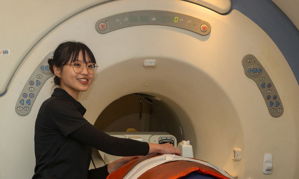
We offer high-field MRI service in Western Australia for small animals, and the first standing MRI service for horses.
The Animal Hospital’s 1.5 Tesla MRI machine is engineered with high density coils, which provide premium quality images with enhanced contrast and reduced blurring. With the ability to image patients from the size of a Chihuahua to a Great Dane with no reduction in quality, patients will be able to undergo same-day MRI imaging in order to achieve better diagnostic outcomes and treatment plans.
In partnership with our radiology specialists at Vet Imaging Specialists, the Animal Hospital is now also accepting referrals for small animal MRI scans.
Additionally, we have recently installed the Hallmarq EQ3 0.27 Tesla scanner, one of only four in Australia. This imaging unit is used predominantly to diagnose causes of lameness of the lower limbs, and along with the new standing CT, completes our Standing Advanced Imaging Suite.
The Advanced Imaging Suite is referral only, and is a facility designed to support the entire equine community in WA. TAHMU is accepting referrals from vets across Western Australia for both MRI and CT cases and are happy to discuss the benefits of each modality, as well as the most appropriate option for clients and horses alike. Racing and Wagering Western Australia are key partners in this project and all active racehorses receive discounted scan fees in recognition of this support.

Radiography
Our facilities include a dedicated large animal radiology suite with wireless digital radiography system and two small animal computed radiology suites.
Highly experienced staff with advanced training in radiology offer opinions on all radiographic studies.
Ultrasound
Our ultrasound systems have an array of different transducers that facilitate examination of a wide range of body systems including the abdomen, thoracic cavity (heart and lungs) and musculoskeletal system of both large and small animals.
Our skilled clinicians also frequently utilise ultrasound guidance to perform precise biopsies of internal structures, in the safest way possible.
Our team
Dr Richardson is a Clinical Veterinary Radiologist, Lecturer in Diagnostic Imaging, a Fellow of the Australian and New Zealand College of Veterinary Scientists in Radiology and Head of the Diagnostic Imaging Section. She is involved in teaching the diagnostic imaging courses in 3rd, 4th, and 5th year of the veterinary program as well as resident training.
She performs imaging procedures within the Animal Hospital at Murdoch University and is involved in the imaging caseload. Her clinical interests include all imaging modalities including radiology; however, has a particular interest in nuclear medicine, cardiac ultrasound, and some advanced imaging techniques.
Dr Josie Faulkner is a Diplomate of the European College of Veterinary Diagnostic Imaging and a Senior Lecturer in Veterinary Diagnostic Imaging (Large Animals) at Murdoch University. She teaches various diagnostic imaging lectures and practicals in the Veterinary Science degree course and is also involved with training residents. Josie enjoys working with all imaging modalities at The Animal Hospital at Murdoch University and is particularly interested in advanced imaging of orthopaedic cases and computed tomography of the head and neck. She is currently finalising her PhD thesis which focuses on magnetic resonance imaging and computed tomography of sagittal groove disease of the equine proximal phalanx.
BVSc MMedVet (Diag Im) Dip ECVDI
Nerissa graduated from the University of Pretoria, South Africa in 2002 after which she spent 4 years working in small animal practice in the United Kingdom, including an internship at a large multidisciplinary referral hospital, Davies Veterinary Specialists. She then returned to South Africa to specialise in Diagnostic Imaging and conducted a parallel European College Diagnostic Imaging residency program. After successful completion of her exams in 2010, she became a South African Veterinary Diagnostic Imaging Specialist and then a Diplomate of the European College of Veterinary Diagnostic Imaging. After her residency she took up a post as Senior Lecturer in Radiology at the University of Pretoria. In July 2012 Nerissa and her family relocated to Perth where she set up the Vet Imaging Specialists business which provides diagnostic imaging services to referring veterinarians as well as The Animal Hospital at Murdoch University. She enjoys teaching and is an accomplished public speaker having extensively presented continuing veterinary education courses and guest lectures. Nerissa has published several papers as well as co-authored and authored book chapters in the BSAVA manuals of canine and feline abdominal and musculoskeletal imaging. Nerissa has a special interest in thoracic imaging, echocardiography, gastrointestinal ultrasonography and MRI but never tires of looking at radiographs!
Dr Ma is a second year Resident of the European College of Veterinary Diagnostic Imaging, having recently attained her Postgraduate Diploma in Veterinary Clinical Studies from Murdoch University. Prior to her time at Murdoch, Doris completed two comprehensive rotating internships at specialist small animal hospitals in New Zealand and Sydney.
At TAHMU, Doris is involved with daily clinical cases that require radiography, ultrasound and advanced imaging, while actively engaging fifth-year students in case discussions. She is enthusiastic about all imaging techniques and continuously seeks to expand her knowledge across all modalities over the next few years of her training at TAHMU.
Pascal graduated from Adelaide University in 2016 and has worked in both general and specialist practice prior to undertaking a diagnostic imaging traineeship in 2024 at Murdoch University. In 2025, Pascal commenced his first year as a Resident of the European College of Veterinary Diagnostic Imaging.
Pascal finds it rewarding how diagnostic imaging is integral in nearly all patients that present through the hospital, and particularly values inter-departmental collaboration in improving patient care. Outside of work he enjoys exploring the great outdoors that WA has on offer with his partner and their Labrador Patrick.
The Diagnostic Imaging team has a dedicated group of registered veterinary nurses, Medical Imaging Technologists (MIT’s) and support staff that are an integral part of the hospital and teaching facility. Our nurses and MIT’s exhibit an expert level of care to their patients and provide support to the veterinary team.

-sitting-in-front-of-tah-signage.tmb-300-sqcrop.jpg?Culture=en&sfvrsn=eb6587e5_1)